Home Science & Medicine Radiation Oncology

Radiation Oncology

Feature articles about advances, challenges, or other aspects of radiation oncology that impacts or has potential impact in thoracic oncology.

The Role for Trimodality Therapy in Stage III NSCLC

By Leah Lawrence

Posted: June 2018

The use of trimodality therapy—the combination of chemoradiotherapy followed by surgery—remains controversial for patients with locally advanced cN2-N3 NSCLC.

According to the National Comprehensive Cancer Network guidelines, definitive chemoradiation therapy is the standard of care for the majority of patients with stage III NSCLC, and trimodality treatment is used only in selected patients with minimal N2 disease.

Results of the Intergroup 0139 study, one of the first randomized studies of concurrent chemoradiotherapy and trimodality approaches, showed that the 5-year progression-free survival was improved in patients who underwent trimodality treatment compared with bimodality therapy alone (hazard ratio = 0.77; 95% CI[0.62, 0.96]); however, this benefit did not translate into an overall survival advantage.1

“In a subset analysis of the study, they showed that patients who underwent a lobectomy did have a survival benefit with trimodality treatment, but this was an unplanned analysis,” said Melissa A.L. Vyfhuis, MD, PhD, of the University of Maryland Medical Center.

The lack of overall survival benefit may have, in part, been due to the trial’s high mortality rate seen with pneumonectomies. Furthermore, in the trial, they used a lower radiation dose of 45 Gy prior to surgical resection to offset the chance of an increase risk in morbidity or mortality associated with higher doses, according to Dr. Vyfhuis. “In the setting of stage III disease, we now know that [45 Gy] is not sufficient for cure,” Dr. Vyfhuis said. Practically speaking, if the tumor is deemed not resectable after such a low dose of radiation, then the patient would have to go back and receive additional radiation therapy, but now having sustained a significant break (typically 1 to 2 weeks) in their radiation treatments, which could affect clinical outcomes.

According to Dr. Vyfhuis, at the University of Maryland, she and her colleagues give a definitive dose of radiation (≥ 60 Gy) with concurrent chemotherapy, even if a patient was scheduled to undergo surgery; however, she acknowledged that not a lot of institutions routinely offer this dose as part of trimodality therapy.

“At University of Maryland, our surgeons have extensive experience operating on patients after the administration of a definitive dose (≥ 60 Gy) of radiation. This has resulted in low rates of postsurgical morbidity and mortality, especially for those patients undergoing a lobectomy,” Dr. Vyfhuis explained.

Dr. Vyfhuis and colleagues recently published the results of a study that showed that trimodality treatment with a radiation dose of at least 60 Gy significantly improved survival and freedom from recurrence in patients with locally advanced NSCLC.2

In our experience, patients who attain mediastinal nodal clearance after neoadjuvant chemoradiation, no matter how bulky or extensive the disease was initially, can benefit from trimodality therapy.
–Melissa A.L. Vyfhuis, MD, PhD


The retrospective analysis included data from 355 consecutive patients with locally advanced NSCLC treated with curative intent between January 2000 and December 2013. Those patients who received trimodality therapy had a significantly longer median survival compared with patients with either unplanned or planned bimodality treatment (59.9 vs. 20.1 vs. 17.3 months, respectively; p < 0.001). The addition of surgery also benefited patients with stage IIIb (p < 0.001) and N3 (p = 0.010) nodal disease, especially when mediastinal nodal clearance was achieved.

“A median survival of approximately 60 months is essentially unheard of in stage III disease,” Dr. Vyfhuis said, adding that as a retrospective study some selection bias may be present. “In our experience, patients who attain mediastinal nodal clearance after neoadjuvant chemoradiation, no matter how bulky or extensive the disease was initially, can benefit from trimodality therapy.”

How Does It Fit?
The current standard of care for patients with stage III NSCLC may soon be changing however, according to Martin J. Edelman, MD, chair of the department of hematology/oncology at Fox Chase Cancer Center, and formerly of the University of Maryland Greenebaum Comprehensive Cancer Center.

In 2017, results of the phase III PACIFIC trial showed that the administration of the anti–PD-1 antibody durvalumab after definitive chemoradiotherapy more than tripled the median progression-free survival compared with chemoradiotherapy followed by placebo (16.8 vs. 5.6 months; p < 0.001).3 The results were presented at the 2017 European Society for Medical Oncology Congress and published in The New England Journal of Medicine. Based on these results, the standard of care today for a patient with locally advanced NSCLC is chemoradiotherapy followed by immunotherapy, according to Dr. Edelman.

“The trial was done predominantly in Europe, a little bit differently than we might have done it in the United States, but results were impressive,” Dr. Edelman said. “We do not yet have overall survival results, but I would be surprised if they do not echo the substantial improvements in progression-free survival that was published.”

The integration of immunotherapy into treatment regimens for patients with stage III disease only further complicates matters. Many questions remain, Dr. Edelman said.

“We still do not know the optimal chemotherapy regimen to use in combination with radiation,” Dr. Edelman said.“We feel following chemoradiotherapy with immunotherapy is good, but do not know if immunotherapy should follow immediately.”

Trimodality care should be restricted to experienced institutions that have high volume and an experienced multimodality team.
–Martin J. Edelman, MD


With so many questions remaining about bimodality therapy, it is hard to know where surgery would fit in.

According to Dr. Edelman, an ideal candidate for trimodality treatment would be someone who is relatively fit, with an otherwise good performance status. Ideally, the patient would require a lobectomy and not a pneumonectomy or another type of complex procedure, and would have mediastinal nodal disease that is not bulky. “Those patients in the correct hands should have a very low operative mortality,” Dr. Edelman said. However, outside of these situations, the standard of care remains bimodality therapy, he added.

“The problem with trimodality studies is how one integrates all three modes of treatment is very difficult, and each study has to be evaluated by itself because no two of them held all features constant,” Dr. Edelman explained.

When he was at the University of Maryland, using a radiation dose of 60 Gy with chemotherapy was feasible. If a patient did not go on to surgery, this meant that the proper definitive radiation dose had been administered. However, this approach may not be feasible in all institutions.

“Trimodality care should be restricted to experienced institutions that have high volume and an experienced multimodality team,” Dr. Edelman said. “Patients who are felt to be suitable for this treatment should be selected prior to initiation of any treatment.” ✦

References:
1. Albain KS, Swann RS, Rusch VR, et al. Radiotherapy plus chemotherapy with or without surgical resection for stage III non-small cell lung cancer. Lancet. 2009;374:379-386.
2. Vyfh uis MAL, Bhooshan N, Burrows WM, et al. Oncological outcomes from trimodality therapy receiving definitive doses of neoadjuvant chemoradiation (≥60 Gy) and factors influencing consideration for surgery in stage III non-small cell lung cancer. Adv Radiat Oncol. 2017;2:259-269.
3. Antonio SJ, Villegas, Daniel D, et al. Durvalumab after chemoradiotherapy in stage III non-small cell lung cancer. N Engl J Med. 2017;377:1919-1929.

Patients with EGFR Mutation Should Postpone Brain Radiation for CNS Metastases: Pro and Con

By Pranshu Mohindra, MD, MBBS, DABR®, Lecia Sequist, MD, and Laurie E. Gaspar, MD, MBA
Posted: December 2017

Since the initial approval of erlotinib, an oral tyrosine kinase inhibitor (TKI), for treatment of patients with previously treated locally advanced or metastatic non-small cell lung cancer (NSCLC), multiple additional treatment agents targeting EGFR mutation are now recommended for use in clinical practice.1 Estimated median survival for patients with previously untreated EGFR-mutant positive (EGFRmt (+)) NSCLC can extend well beyond 2 years; however, this increase in longevity has been linked with an increased incidence of brain metastases (BM).2,3 While whole-brain radiotherapy (WBRT) was previously considered the standard of practice, the concern for neurocognitive side effects has led to the decreased use of WBRT in favor of stereotactic radiosurgery (SRS), as supported by phase-III trials that do not demonstrate a detriment in survival.4-6

EGFRmt (+) NSCLC provides a unique therapeutic setting where even with a diagnosis of BM, extended survival may be seen.7 A debate was conducted at the recently concluded 2017 IASLC meeting in Chicago, reviewing the pros and cons of withholding radiation therapy in patients with EGFRmt (+)-NSCLC diagnosed with BM. Key highlights from the debate presented by Dr. Lecia Sequist (Pro) and Dr. Laurie Gaspar (Con) are excerpted below.

Pro: Dr. Lecia Sequist 

1. A multi-institutional retrospective study evaluated upfront radiation (WBRT or SRS) approaches versus upfront EGFR-directed therapy approaches for these patients and showed a significant detriment in overall survival (OS) by delayed use of either of the radiation options.8 However, there are limitations in this experience, with other literature review showing mixed results:

• The presence of central nervous system (CNS)-only disease in 76% of the patients likely biased the outcomes in favor of upfront radiation.
• Also, SRS was planned only at intracranial progression, not as planned consolidation of residual disease.
• In a literature review of other institutional experiences, mixed results were observed, with 2 other studies showing survival advantage with use of radiation, 1 study showing survival advantage with TKI alone, and 3 other studies showing no significant differences, although a trend of a 4- to 7-month survival improvement with radiation was noted.

2. All published studies to date used erlotinib- or gefitinib-based therapy, which have demonstrated less CNS penetration compared to newer EGFR TKIs. Outcomes are superior in the osimertinib era.

• AURA 2 study, a phase II study evaluating use of AZD9291 (osimertinib) in EGFR and T790M mutation positive tumors after previous EGFR TKI therapy, demonstrated a 54% overall response rates (ORR) within the brain (2016 World Conference on Lung Cancer, Vienna, Austria).
• The phase I BLOOM study evaluated the use of osimertinib in patients with EGFRmt (+) leptomeningeal disease. Among 21 patients, efficacy assessments confirmed radiological response in 7 and cytological CSF clearance in 2 patients (2016 ASCO Annual Meeting, Chicago, US).
• AURA 3 study demonstrated that the ORR in brain with osimertinib in comparison with chemotherapy in patients with progression after first-line TKI therapy were 70% vs. 31%, p = 0.015 (2017 ASCO Annual Meeting, Chicago, USA). In a follow-up detailed report of this phase 3 study, analysis of 144 patients with T790M-positive advanced NSCLC who develop BM demonstrated a significant improvement in progression-free survival (PFS) favoring osimertinib as against platinum-pemetrexed chemotherapy doublet: 8.5 months vs 4.2 months (HR, 0.32; 95% CI, 0.21 to 0.49).9
• In the FLAURA study comparing osimertinib to standard therapy (erlotinib or gefitinib) in EGFRmt (+) -NSCLC, in patients with BM (n = 116), the median PFS with osimertinib versus standard therapy was 15.2 months vs. 9.6 months (HR, 0.47; 95% CI, 0.30-0.74; P = 0.0009). The rates of CNS progression were 6% versus 15%, respectively (2017 ESMO Congress, Madrid, Spain).

Bottom line: Given the risk of radionecrosis or steroid dependence from SRS or cognitive decline from WBRT, and in light of particularly encouraging outcomes from recent studies evaluating osimertinib in BM, I support use of upfront systemic therapy to offer patients an opportunity for response and thereby delay the risk of side effects from the use of radiation therapy. I also favor consideration of SRS to any significant residual CNS lesions after initial response to TKIs, a sequence and therapeutic strategy employed increasingly at most academic centers, an approach that has not been permitted in most published studies.

Con: Dr. Laurie E. Gaspar 

1. The prognosis of EGFRmut (+) BM and the time to salvage SRS/ WBRT is not as good as perceived, especially if BM occur while on TKI.

• In the Massachusetts General Hospital experience, patients with EGFRmt (+) or ALK translocation who developed BM in the setting of prior TKI therapy had worse OS than those not on TKI prior to the BM diagnosis (median OS 9m vs. 19.6 m, p < 0.001).10 Further, after cranial radiotherapy, EGFR mutation status did not impact OS.10
• In a multi-institutional retrospective database, median OS after diagnosis of BM for EGFRmt patients was 23 months (17 months for TKI treated versus 30 months for TKI-naive patients, p < 0.01). When time-dependent analysis was performed, extended survival associated with EGFRmt (+) NSCLC was only noted in TKI-naive patients relative to those who developed BM while on TKI therapy.11
• Even on the AURA 3 study, despite the 70% ORR, median PFS for patients whose disease had progressed on first-line TKI and develop CNS disease was only 8.5 months.9
• In a phase-II Japanese study using gefitinib in EGFRm-NSCLC with brain metastases, despite a 87.8% ORR, the median time on gefitinib was only 10.6 months with intracranial progression being the most common cause of withdrawal.12

2. While neurocognitive effects following WBRT are well known, there are no comparable data on the neurocognitive effects of TKI.

3. In afatinib-treated patients in the LUX-Lung 3 and LUX-Lung 6 trials, the benefit of afatinib appeared higher in patients with prior WBRT with median PFS in entire cohort ranging from 8.2 to 11 months.13

4. Results reported in the study by Magnuson et al are compelling across all prognostic subpopulations.8 This multi-institutional retrospective study evaluated upfront radiation (WBRT or SRS) approaches versus upfront EGFR-directed therapy approaches for these patients and showed a significant detriment in OS by delaying implementation of either of the radiation options. Key findings are:

• Median OS for upfront-SRS, upfront-WBRT and upfront EGFR-TKI, with SRS or WBRT at intracranial progression, were 46, 30, and 25 months, respectively, p < 0.001.
• In both radiation cohorts, 50% of patients were symptomatic at the time WBRT was initiated, compared to only 12% of patients in the EGFR-TKI cohorts.
• Even after controlling for variables that constitute the Disease Specific Graded Prognostic Assessment (DS-GPA) score7 and the EGFRm status, upfront SRS was independently associated with improved OS relative to EGFR-TKI and delayed radiation (adjusted HR, 0.39; 95% CI, 0.26 t 0.58, p < 0.001).
• Prior EGFR-TKI use and EGFR-TKI resistance mutations were exclusions, thereby suggesting the benefit of radiation was even more pronounced in the better prognostic group.

5. The biggest concern is the overall quality of life for patients with BM and the symptoms and sequelae from the metastatic intracranial burden. Bottom line: The argument for upfront radiation is especially strong for SRS, as opposed to WBRT, so why wait and let a BM get larger or more symptomatic, and not be amenable to SRS? At the University of Colorado, these patients are given upfront SRS if possible, and then proceed to TKI. If SRS is not thought to be reasonable, then the TKI is started and SRS or WBRT is deferred until progression.

Audience response: There was a lively discussion followed by an informal vote that (predictably) declared no clear winner. ✦

References
1. NCCN Clinical Practice Guidelines in Oncology (NCCN Guidelines) Non-Smal Cell Lung Cancer version 9.2017, Natl. Compr. Cancer Network. (2017). https://www.nccn.org/professionals/physician_gls/pdf/nscl.pdf (accessed October 21, 2017).
2. Jackman DM, Miller VA, Cioffredi LA, et al. Impact of epidermal growth factor receptor and KRAS mutations on clinical outcomes in previously untreated non-small cell lung cancer patients: results of an online tumor registry of clinical trials. Clin Cancer Res. 2009; 15:5267-5273.
3. Shin DY, Na II, Kim CH, Park S, Baek H, Yang SH. EGFR mutation and brain metastasis in pulmonary adenocarcinomas. J Thorac Oncol. 2014; 9:195-199.
4. Chang EL, Wefel JS, Hess KR, et al. Neurocognition in patients with brain metastases treated with radiosurgery or radiosurgery plus whole-brain irradiation: A randomised controlled trial. Lancet Oncol. 2009; 10:1037–1044.
5. Brown PD, Jaeckle K, Ballman KV, et al. Effect of radiosurgery alone vs radiosurgery with whole brain radiation therapy on cognitive function in patients with 1 to 3 brain metastases: A randomized clinical trial. JAMA. 2016; 316:401–409.
6. Brown PD, Ballman KV, Cerhan JH, et al. Postoperative stereotactic radiosurgery compared with whole brain radiotherapy for resected metastatic brain disease (NCCTG N107C/CEC•3): A multicentre, randomised, controlled, phase 3 trial. Lancet Oncol. 2017; 18:1049–1060.
7. Sperduto PW, Yang TJ, Beal K, et al. Estimating survival in patients with lung cancer and brain metastases: An update of the graded prognostic assessment for lung cancer using molecular markers (Lung-molGPA). JAMA Oncol. 2017; 3:827–831.
8. Magnuson WJ, Lester-Coll NH, Wu AJ,et al. Management of brain metastases in tyrosine kinase inhibitor-naïve epidermal growth factor receptormutant non-small-cell lung cancer: A retrospective multi-institutional analysis. J Clin Oncol. 2017; 35:1070–1077.
9. Mok TS, Wu YL, Ahn MJ, AURA3 Investigators, et al., Osimertinib or Platinum-Pemetrexed in EGFR T790M-Positive Lung Cancer. N Engl J Med. 2017; 376:629–640.
10. Mak KS, Gainor JF, NiemierkoA, et al. Significance of targeted therapy and genetic alterations in EGFR, ALK, or KRAS on survival in patients with nonsmall cell lung cancer treated with radiotherapy for brain metastases. Neuro Oncol. 2015; 17:296–302.
11. Sperduto PW, Yang TJ, Beal K, et al. The effect of gene alterations and tyrosine kinase inhibition on survival and cause of death in patients with adenocarcinoma of the lung and brain metastases. Int J Radiat Oncol Biol Phys. 2016; 96:406–413.
12. Iuchi T, Shingyoji M, Sakaida T, et al. Phase II trial of gefitinib alone without radiation therapy for Japanese patients with brain metastases from EGFR-mutant lung adenocarcinoma. Lung Cancer. 2013; 82:282–287.
13. Schuler M, Wu YL, Hirsh V, et al. First-line afatinib versus chemotherapy in patients with non-small cell lung cancer and common epidermal growth factor receptor gene mutations and brain metastases. J Thorac Oncol. 2016; 11:380–390.

Utility and Controversies Surrounding Use of New Techniques in Radiotherapy

By Suresh Senan, MRCP, FRCR, PhD
Posted: December 2017

Improved delivery of radiotherapy has contributed to the recent changes in guidelines for patients with lung cancer. The clinical impact of some technical advances has been modest, but the cumulative effect of many developments has led to more precise treatment delivery, and has increased the confidence of clinicians to adopt new techniques. The impact of new technology is best illustrated by referring to selected clinical stages.

Early-stage Peripheral NSCLC

The poster child of new technology is image-guided stereotactic ablative radiotherapy (SABR or SBRT), which is a technique for delivering high-dose, high-precision radiation. In early-stage peripheral non-small cell lung cancer (NSCLC), 5-year in-field tumor control rates of 90% have been reported, and the introduction of SABR has been associated with improved cure rates at the population level. The superiority of SABR over conventional radiotherapy has been established in two randomized trials, results from one of which was presented at the IASLC 18th World Conference on Lung Cancer [Ball D, WCLC 2017]. SABR resulted in superior freedom from local failure (HR = 0.29, 95% CI 0.130, 0.662, P=0.002) and also longer overall survival (HR = 0.51, 95% CI 0.51, 0.911, P=0.020).

Minimal requirements for SABR planning and delivery were recently published by the European Society for Radiotherapy and Oncology, and include use of 4-D planning computed tomography (CT) scan and a standard linear accelerator with a cone-beam CT scan. Newer developments include the clinical introduction of magnetic resonance imaging-guided SABR, which is undergoing evaluation in high-risk tumors such as centrally located NSCLC. The use of protons (charged particles) was considered previously to be a promising development for treating lung tumors, due to its ability to decrease radiation doses to surrounding organs. However, the options for managing tumor motion at current proton centers is generally inferior to that available on a standard linear accelerator. At present, there are limited prospective data supporting a role for protons in early-stage NSCLC.

Locally Advanced Lung Cancer

Since the introduction of CT-based treatment planning and improved set-up protocols, concurrent chemo-radiation (CT-RT) has become established as the standard of care for most fit patients who present with locally-advanced NSCLC. Patients undergoing standard concurrent CT-RT to 60 Gy in the RTOG 0617, study achieved 5-year overall and progression free rates of 32% and 18%, respectively.1 The median overall survival of 28.7 months in this trial has also established a new benchmark. The role of intensity modulated radiotherapy (IMRT) remains a topic of debate, particularly as population studies suggest that benefits of IMRT are limited to larger central (T3-4) tumors. Results of RTOG 0617 support use of IMRT in locally-advanced NSCLC as this reduces rates of radiation pneumonitis, and was associated with a better quality of life in the first 12 months post CT-RT.

Some, but not all studies, suggested that delivery of higher radiation doses to the heart are associated with a poorer overall survival. However, other studies suggest that delivered heart doses may be a surrogate for other prognostic factors in stage III NSCLC, such as the extent and location of mediastinal nodal involvement, especially subcarinal nodes, rather than an independent predictor of outcome. The increased cardiac toxicity has also been correlated with the use of doses higher than the standard 60 Gy for CT-RT, and with schemes using non-standard fractionation schemes (>2 Gy, once daily).

Proponents of proton radiation have therefore advocated its use as a means to decrease both lung and cardiac toxicity. The number of proton centers worldwide is increasing in a roughly exponential fashion, with approximately 20 operating centers, and 53 in development. However, little consensus has been reached for using proton therapy for common types of cancer in adults. A prospective randomized trial led by the MD Anderson Cancer Center compared the use of IMRT versus proton therapy in locally advanced NSCLC, and reported no differences in treatment failures, which were defined as either grade ≥3 pneumonitis or local failure at 1 year.2 Proponents of protons have argued that the use of a newer delivery technique (intensity-modulated proton therapy) will improve outcomes, and the results of ongoing comparative trials are awaited. Treatment of mobile tumors in the lung and liver using protons remains a challenge, and only a minority (27%) of European proton and carbon ion therapy centers currently treat such tumors.3

Changing Paradigms in Locally Advanced Lung Cancer

The published results of the PACIFIC trial have called into question the previous focus on delivery of ever higher radiation doses. PACIFIC evaluated consolidation durvalumab or placebo every 2 weeks for a year, following CT-RT in patients with unselected stage III NSCLC after concurrent CT-RT to a dose of 54-66 Gy.4 Consolidation durvalumab resulted in an increase in median PFS to 16.8 versus 5.6 months (HR 0.52) with the differences in PFS sustained at 12- and 18-month landmarks. Durvalumab also resulted in a superior median time to death or distant metastases (23.2 vs 14.6 months; P < .001), and little increase in grade 3/4 treatment related toxicity. The above findings indicate that the addition of durvalumab consolidation to standard radiation doses combined with two cycles of platinum-containing chemotherapy, is sufficient to improve both local and distant tumor control. This, in turn, raises the question whether the focus of radiation research should simply be to limit radiation doses to 60 Gy, while further optimizing the integration of immune-oncology and other systemic approaches into CT-RT of stage III NSCLC. ✦

References
1. Bradley JD, Hu C, Komak RU, et al. Long-term results of RTOG 0617: A randomized phase 3 comparison of standard dose versus high dose conformal chemoradiation therapy +/- cetuximab for stage III NSCLC. J Am Coll Radiol. 2017:99, S105 (suppl)
2. Liao Z, Lee J, Komaki R, Gomez D, O’Reilly M, Allen P, et al. Bayesian randomized trial comparing intensity modulated radiation therapy versus passively scattered proton therapy for locally advanced non-small cell lung cancer. J Clin Oncol. 2016;34 suppl 15:8500.
3. Weber DC, Abrunhosa-Branquinho A, Bolsi A, et al. Profile of European proton and carbon ion therapy centers assessed by the EORTC facility questionnaire. Radiother Oncol. 2017;124:185-189.
4. Scott J. Antonia SJ, Villegas A, Davey Daniel D, et al. Durvalumab after chemoradiotherapy in stage III non–small-cell lung cancer. N Engl J Med. 2017 Sep 8. doi: 10.1056/ NEJMoa1709937. [Epub ahead of print]

Proton-Beam Therapy Versus Photon-Beam Therapy: The Debate Continues

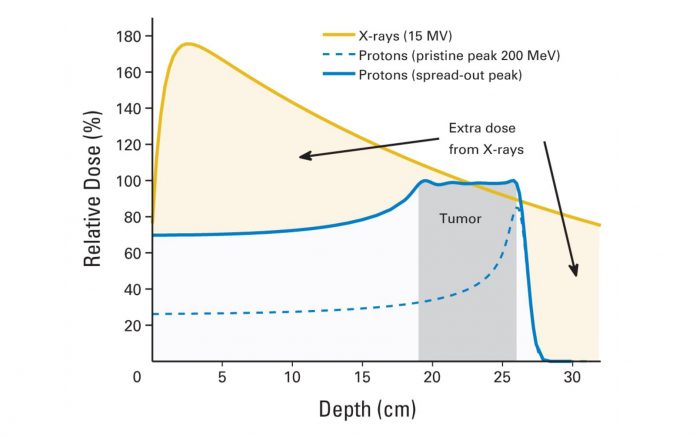
Comparison of relative depth dose distributions of photons versus protons. Source: J Clin Oncol. 2014 Sep 10;32(26):2855-63.

By Cynthia L. Kryder, MS, CCC-Sp
Posted: October 2017

For patients who present with inoperable, locally advanced lung cancer, photon-based chemoradiation remains the standard of care. Despite advanced radiation-delivery techniques, such as multi-leaf collimators, intensity-modulated radiotherapy (IMRT), and imageguided radiotherapy (IGRT), radiation oncologists continue to explore ways to extend the ALARA principle, that is, the desire to deliver tumoricidal radiation doses to intended targets while minimizing the radiation doses to adjacent healthy tissues. This has led radiation oncologists to investigate the potential of proton beam radiation therapy. In patients with non-small cell lung cancer (NSCLC), proton-beam therapy may enable safe dose escalation while sparing chest organs at risk and simultaneously maintaining adequate target coverage. In so doing, the collateral damage of standard radical thoracic radiotherapy can, theoretically, be mitigated.

Photons Versus Protons

Although the therapeutic index of modern, highly conformal photon radiotherapy has increased, the physics of photons make it impossible to avoid the exit dose downstream from the target, which is a physical limitation of the photon beam. In comparison, protons travel through tissue quickly and stop abruptly when reaching tissues at a very specific depth. Unlike photons, which deposit their radiation doses close to their entrance into the body, protons deposit most of their energy at the end of their paths, in a phenomenon known as the Bragg peak, the point at which the majority of energy deposition occurs. Before the Bragg peak, the deposited dose is about 30% of the Bragg peak maximum dose. Thereafter, the deposited dose falls to practically zero, yielding a nearly nonexistent exit dose. The integral dose with proton therapy is approximately 60% lower than any photon-beam technique.1 Thus, proton therapy delivers radiation to tumors and areas in very close proximity, decreasing integral radiation dose to normal tissues and theoretically avoiding collateral damage.

Despite these potential advantages, a fundamental issue with protons is the ability to stop the proton at the tumor. As any external beam travels through the body toward its target, it passes through tissues of different densities. Protonbeam therapy is much more sensitive to tissue density than photon therapy. Likewise, at greater depths the lateral margins of the proton beam become less sharp due to considerable scattering.2 Any change in tissue composition, such as organ motion, lung expansion, or alteration in bone position from one treatment to the next, can affect target coverage and dose to surrounding structures. To account for tissue heterogeneity and to reduce the potential for tumor underdosing, radiation oncologists often add a margin of uncertainty, meaning that the beam is designed to overshoot the target to guarantee good coverage.3 This could, however, negate the tissue-sparing advantage of proton-beam therapy and/or dilute its therapeutic effects.

Another difference between photon beam therapy and proton-beam therapy is the expense. Proton-beam therapy is an expensive technology. Including a cyclotron, multistory gantries, and several treatment rooms, the average cost for a proton facility ranges between US$140 million and US$200 million.

Assessing the Clinical Advantage of Proton-Beam Therapy

Given its lower integral dose and steeper dose gradient, proton therapy is an appealing therapeutic option. However, dosimetry advantages alone will not be enough to convince payors and patients to adopt this costly technology. Proton beam therapy must demonstrate a measurable clinical advantage when compared with standard photon therapy.

Clinical trials are underway to do just that. Zhongxing Liao, MD, of the Department of Radiation Oncology at the University of Texas MD Anderson Cancer Center, is the principal investigator of a multi-center, prospective, randomized phase III trial that will compare overall survival after photon versus proton chemoradiotherapy in patients with unresectable locally advanced NSCLC.4 This randomized trial will compare the overall survival (OS) in patients with stage II-IIIB NSCLC after image-guided, motion-managed photon radiotherapy (Arm 1) or after image-guided, motion-managed proton radiotherapy (Arm 2), both given with concurrent platinum-based chemotherapy. A total of 560 patients are expected to be enrolled. The primary endpoint is OS; secondary endpoints include 2-year progression-free survival, adverse events, quality of life, cost-effectiveness, and changes in pulmonary function.

A second ongoing trial seeks to determine whether the dose of radiation to the tumor, but not the surrounding healthy tissue, could be increased by using IMRT or intensity-modulated proton beam therapy (IMPT).5 In phase I of the study, investigators will identify the maximum tolerated dose (MTD) of IMPT and IMRT. In phase II, researchers will compare the efficacy of IMPT and IMRT when both treatments are combined with standard chemotherapy. The primary outcome measure is MTD; the secondary outcome measure is progression-free survival.

Future Outlook

The ability of proton-beam therapy to precisely target tumors and spare underlying tissues from radiation exposure in patients with a variety of cancers has already been demonstrated. Exactly if and how proton-beam therapy fits into the treatment of patients with lung cancer remains to be determined. Harnessing the power of proton-beam therapy in the treatment of NSCLC may be challenging given that protons must be delivered to the lungs, which are targets in motion that are surrounded by tissues of different densities. Future studies will need to assess not only side effects and outcomes, but they will also need to provide data to support the development of dose algorithms and motion-management techniques.

Given the capital investment and operating costs associated with protonbeam therapy, examining the economic advantages and liabilities of this new technology is necessary. Clear data about its cost effectiveness based on different clinical and treatment scenarios will enable providers, payors, and patients to make informed decisions about treatment. ✦

Expert Comment
The photon versus proton conundrum continues in the latter part of 2017, and it now must evolve in the context of promising new data with immune enabling drugs such as checkpoint inhibitors. Personally, I believe it is unlikely that further dose escalation to the target area will result in significant benefits in local control and overall survival from a radiobiologic perspective despite potential advantages in dose deposition by proton therapy, so newer directions are needed. From a cost perspective, is a 140-200 million monetary outlay for protons the way to get us to the promised land? Or will molecular and immunological discoveries offer the best avenue for success? Perhaps radiation, whether through protons or photons, will be the match rather than the flame for immune enabling drugs; therefore, dose escalation may be less important. Building on the theme of potential clinical advantages between photon or proton intensity modulated therapy, the question is whether less integral dose scatter within normal tissue with the use of protons will result in less chronic immunosuppression and thus potentiate checkpoint inhibition over photon irradiation. This is an amazing opportunity to study the changes in lymphocyte:neutrophil ratios during and after treatment. The bar has jumped with the anticipated results of the PACIFIC trial in locally advanced NSCLC, and we must jump with it. —David Raben, MD

References

1. Mitin T, Zietman A. Promises and pitfalls of heavyparticle therapy. J Clin Oncol. 2014;32:2855-2863.
2. Goitein M. Magical protons? Int J Oncol Biol Phys. 2008;70:654-656.
3. Paganetti H. Range uncertainties in proton therapy and the role of Monte Carlo simulations. Phys Med Biol. 2012;57:R99–R117.
4. ClinicalTrials.gov [website]. Comparing photon therapy to proton therapy to treat patients with lung cancer. Last updated June 10, 2016. https:// clinicaltrials.gov/ct2/show/NCT01993810. Accessed July 24, 2017.
5. ClinicalTrials.gov [website]. Intensity-modulated scanning beam proton therapy (IMPT) with simultaneous integrated boost (SIB). Last updated July 22, 2016. https://clinicaltrials.gov/ct2/show/NCT01629498. Accessed July 24, 2017.

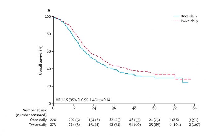
CONVERT: An International Randomized Trial of Concurrent Chemo-Radiotherapy Comparing Twice-Daily and Once-Daily Radiotherapy Schedules in Patients with Limited-Stage Small Cell Lung Cancer and Good Performance Status

Source: http://www.thelancet.com/journals/lanonc/article/PIIS1470-2045(17)30318-2/fulltext

By Prof. Corinne Faivre-Finn

The optimal timing and schedule of thoracic radiation in the management of limited-stage small cell lung cancer (LS-SCLC) continues to provoke debate. Since the publication of Intergroup 0096 in 1999, there had been controversy about the standard chemo-radiotherapy (cCTRT) regimen in LS-SCLC.1 Although twice-daily (BD) radiotherapy (RT) was associated with a higher survival compared to once-daily (OD) RT, concerns regarding toxicity (i.e., a third of the patients developing grade 3 or more esophagitis), together with logistical issues in the delivery of BD RT and criticism about the low dose of RT used in the control arm of Intergroup 0096, led to the limited adoption of this regimen in routine practice.2

The CONVERT trial is the first multicenter, international, randomized phase III trial aiming to establish a standard chemo-radiotherapy regimen in LS-SCLC. It is the largest-ever trial completed in this group of patients. We reported the trial results at the 2016 Annual Meeting of the American Society of Clinical Oncology.3

In CONVERT, patients with LS-SCLC were randomized 1:1 to receive either 45 Gy in 30-BD fractions over 3 weeks or 66 Gy in 33-OD fractions over 6.5 weeks, starting on day 22 of cycle 1 chemotherapy (four to six cycles of cisplatin and etoposide, according to investigator’s prespecified choice), followed by prophylactic cranial irradiation, if indicated. RT was planned using either 3-D conformal or intensity-modulated radiation therapy (IMRT). The primary endpoint of the study was 2-year survival and all analyses were by intention to treat. The study enrolled 547 patients recruited from 73 centers in seven European countries and Canada between 2008 and 2013.

Patient characteristics were well balanced in both arms of the study. Median age was 63 years (15% were older than 70 years), almost 50% were female, and the majority of patients had a performance status of 0 or 1 and were ex-smokers or current smokers.

At a median follow-up of 45 months, 2-year survival was 56% compared to 51%, and median overall survival was 30 months compared with 25 months in the BD RT and the OD RT arms, respectively, a difference that did not prove to be statistically significant. Furthermore, no statistically significant differences between the two groups were reported in terms of local or metastatic disease progression.

Acute toxicity rates were not significantly different between the two groups, with the exception of more neutropenia in patients treated with BD RT. Nor was there any difference in terms of acute esophagitis or pneumonitis. There was one death in the BD group and two in the OD group due to radiation pneumonitis. Few patients developed severe late toxicity.

In conclusion, OD RT did not result in a superior survival or worse toxicity than BD RT. The survival for both regimens was higher than previously reported, possibly due to more frequent PET/CT staging, and radiation toxicities were lower than expected, likely due to using modern RT techniques. The implications of CONVERT are important. The OD arm did not show superior survival as originally postulated. Because CONVERT was not an equivalency trial, the only study to date that has shown superiority for one RT regimen over another in LS-SCLC was the Intergroup 0096 trial; because there were no major differences in toxicity, 45 Gy in 30-BD fractions should continue to be regarded as standard of care. However, OD RT at a dose of 66 Gy in 33 fractions can certainly be considered an alternative regimen if 45 Gy in 30-BD fractions cannot be delivered due to patients’ choice, departmental logistics, or other factors.

References
1. Turrisi AT, Kim K, Blum R, Sause WT, Livingston RB, Komaki R, et al. Twice-daily compared with once-daily thoracic radiotherapy in limited small-cell lung cancer treated concurrently with cisplatin and etoposide. N Engl J Med. 1999; 340:265–271.

2. Komaki R, Khalid N, Langer CJ, et al. Penetration of recommended procedures for lung cancer staging and management in the United States over 10 years: a quality research in radiation oncology survey. Int J Radiat Oncol Biol Phys. 2013; 85:1082-1089.

3. Faivre-Finn C, Snee M, Ashcroft A, et al. CONVERT: An international randomised trial of concurrent chemo-radiotherapy (cCTRT) comparing twice-daily (BD) and once-daily (OD) radiotherapy schedules in patients with limited stage small cell lung cancer (LS-SCLC) and good performance status (PS). J Clin Oncol. 2016; 34 (15) suppl: 8504.

Meeting News